Embark on a captivating journey into the realm of microbiology with Simple Stains Data Sheet 3-4, your ultimate guide to understanding the fundamentals of staining techniques. From the purpose and usage of simple stains to their applications in the field, this data sheet unravels the mysteries of bacterial identification, making complex concepts accessible and engaging.
Delve into the intricacies of Gram staining, a cornerstone of microbiology, and uncover the role of each reagent in revealing the hidden characteristics of bacteria. Step-by-step instructions guide you through the staining process, ensuring accurate and reliable results.
Simple Stains Data Sheet 3-4 Overview

Simple Stains Data Sheet 3-4 provides a comprehensive guide to the preparation and use of simple stains in microbiology. Simple stains are essential tools for visualizing microorganisms under a microscope, and this data sheet provides detailed instructions for using the most common types of simple stains.
Types of Simple Stains
The data sheet describes three main types of simple stains:
- Methylene Blue: A basic dye that stains bacterial cells blue.
- Crystal Violet: A basic dye that stains bacterial cells purple.
- Safranin: A basic dye that stains bacterial cells red.
Applications of Simple Stains
Simple stains are used in a variety of applications in microbiology, including:
- Identification of bacteria: Simple stains can be used to differentiate between different types of bacteria based on their size, shape, and staining characteristics.
- Detection of microorganisms: Simple stains can be used to detect the presence of microorganisms in clinical specimens, such as blood, urine, and sputum.
- Study of bacterial morphology: Simple stains can be used to study the morphology of bacteria, including their shape, size, and arrangement.
Staining Procedures

Staining procedures are essential techniques used to enhance the visibility and differentiation of microorganisms under a microscope. Gram staining is a widely used staining technique that classifies bacteria into two distinct groups: Gram-positive and Gram-negative. This classification is based on the bacteria’s cell wall structure and its reaction to the Gram stain.
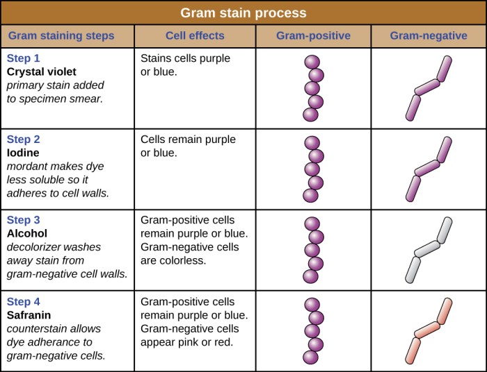
The Gram staining procedure involves the sequential application of various reagents, each playing a specific role in the staining process.
Gram Staining Procedure
- Crystal Violet Application:A crystal violet solution is applied to the bacterial smear, allowing it to penetrate the cell walls of both Gram-positive and Gram-negative bacteria.
- Iodine Application:Gram’s iodine solution, a mordant, is added to the smear, forming a complex with crystal violet within the cells.
- Alcohol Decolorization:Ethanol or acetone is used as a decolorizing agent. Gram-negative bacteria lose the crystal violet-iodine complex due to their thinner cell walls, while Gram-positive bacteria retain the complex.
- Counterstaining:A safranin or fuchsin solution is applied as a counterstain, staining the decolorized Gram-negative bacteria.
Expected Results and Interpretation
After Gram staining, Gram-positive bacteria appear purple or blue, while Gram-negative bacteria appear pink or red. This distinction arises from the differences in their cell wall structures. Gram-positive bacteria have a thick peptidoglycan layer in their cell walls, which retains the crystal violet-iodine complex during the decolorization step.
In contrast, Gram-negative bacteria have a thinner peptidoglycan layer and an outer membrane, which allows the crystal violet-iodine complex to be removed during decolorization, resulting in counterstaining by safranin.
Gram staining is a crucial technique for identifying and classifying bacteria, aiding in their diagnosis and treatment.
Microscopic Examination

Microscopic examination is a fundamental step in simple staining techniques. It allows us to visualize the stained microorganisms and observe their morphology, arrangement, and other characteristics.
Preparing and Observing Stained Slides
To prepare a slide for microscopic examination, the stained slide is placed on the microscope stage and secured with the slide holder. The condenser is adjusted to optimize the illumination, and the objective lens is selected based on the desired magnification.
Once the slide is prepared, the microscope is focused by adjusting the coarse and fine focus knobs. The coarse focus is used to bring the slide into approximate focus, while the fine focus is used for precise adjustment. Proper lighting is essential for clear observation.
The light intensity and condenser position should be adjusted to provide sufficient illumination without overexposing the specimen.
Importance of Proper Lighting and Focusing Techniques
Proper lighting and focusing techniques are crucial for accurate microscopic examination. Inadequate lighting can make it difficult to visualize the stained microorganisms, while excessive lighting can damage the specimen. Proper focusing ensures that the image is sharp and clear, allowing for detailed observation of the microorganisms’ morphology and arrangement.
Data Interpretation
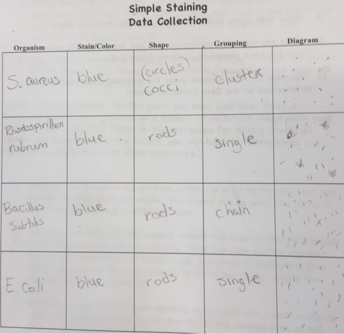
Interpreting simple staining results involves examining the stained cells under a microscope and analyzing their morphological characteristics, such as shape, size, and arrangement.
The primary objective of simple staining is to differentiate between bacteria based on their cell wall composition, which can be Gram-positive or Gram-negative. Gram-positive bacteria retain the crystal violet stain, appearing purple or blue, while Gram-negative bacteria lose the crystal violet stain and instead take up the counterstain, appearing pink or red.
Simple stains data sheet 3-4 is an excellent resource for understanding the various types of simple stains. It provides detailed information on their preparation, uses, and limitations. Just like the classic Grapes of Wrath Chapter 9 , this data sheet is a valuable tool for anyone interested in microbiology.
It helps you identify and differentiate different types of bacteria, which is crucial for accurate diagnosis and treatment.
Morphological Characteristics of Bacteria Revealed by Simple Stains
- Shape:Bacteria can be spherical (cocci), rod-shaped (bacilli), or spiral-shaped (spirilla).
- Size:Bacteria vary in size, typically ranging from 0.5 to 5 micrometers in diameter.
- Arrangement:Bacteria can be found singly, in pairs (diplococci), in chains (streptococci), or in clusters (staphylococci).
Limitations and Potential Errors in Simple Staining Interpretation
- Autolysis:Over-decolorization can lead to false-negative results, as the cell wall may be damaged, causing the Gram-positive bacteria to lose the crystal violet stain.
- Over-staining:Excessive staining can make it difficult to distinguish between Gram-positive and Gram-negative bacteria.
- Contamination:The presence of other microorganisms or debris can interfere with the staining process and lead to misinterpretation.
Reporting and Documentation

Accurate and clear documentation of simple staining results is crucial for effective communication and interpretation. This section provides guidelines for reporting the findings, emphasizing the importance of precision and clarity.
Guidelines for Reporting
- Describe the specimen:Specify the type and source of the specimen (e.g., blood smear, tissue section).
- State the staining technique:Indicate the specific simple staining method used (e.g., Gram stain, methylene blue stain).
- Report the results:Describe the staining characteristics of the microorganisms or cells, including shape, size, color, and any other relevant observations.
- Include negative and positive controls:Report the results of any negative and positive controls used to validate the staining procedure.
- Provide interpretation:Based on the staining results, provide an interpretation of the findings, indicating the presence or absence of microorganisms or specific cell types.
Importance of Accurate Documentation
Accurate and clear documentation ensures:
- Reliable interpretation:Allows healthcare professionals to make informed decisions based on the accurate reporting of staining results.
- Effective communication:Facilitates the sharing of information among healthcare professionals involved in patient care.
- Quality control:Enables the monitoring and improvement of staining procedures, ensuring consistent and reliable results.
- Legal and ethical considerations:Provides a record of the staining process and results for legal and ethical purposes.
Use of Tables and Images, Simple stains data sheet 3-4
Tables and images can enhance the clarity and impact of simple staining reports:
- Tables:Can be used to summarize the staining characteristics of different microorganisms or cell types.
- Images:Can provide visual evidence of the staining results, allowing for more detailed analysis and interpretation.
Quality Control: Simple Stains Data Sheet 3-4

Quality control is crucial in simple staining to ensure accurate and reliable results. It involves following standardized procedures, monitoring the staining process, and identifying and correcting potential errors.
Steps involved in quality control include:
- Using reagents and materials from reputable manufacturers and checking their expiration dates.
- Calibrating and maintaining equipment such as microscopes and staining jars.
- Monitoring the staining time and temperature to ensure consistency.
- Examining stained slides under a microscope to assess staining quality and identify any artifacts or errors.
Potential sources of error in simple staining include:
- Incorrect reagent concentrations or staining times.
- Contamination of reagents or slides.
- Improper handling of slides during staining.
- Suboptimal microscope settings.
Minimizing errors involves adhering to standardized protocols, using high-quality reagents and equipment, and training personnel on proper staining techniques.
Commonly Asked Questions
What is the purpose of Simple Stains Data Sheet 3-4?
Simple Stains Data Sheet 3-4 provides a comprehensive guide to simple staining techniques, including their purpose, usage, and applications in microbiology.
What types of simple stains are included in this data sheet?
The data sheet covers various simple stains, including Gram staining, which is a fundamental technique for differentiating bacteria based on their cell wall structure.
How do I interpret the results of simple staining?
The data sheet provides clear guidelines on interpreting staining results, explaining the different morphological characteristics of bacteria revealed by simple stains.