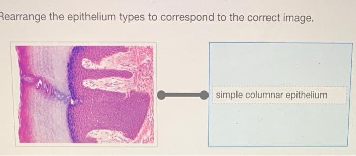
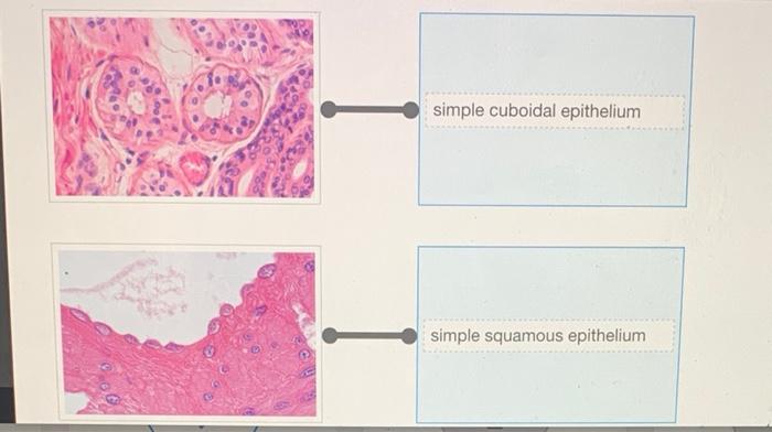
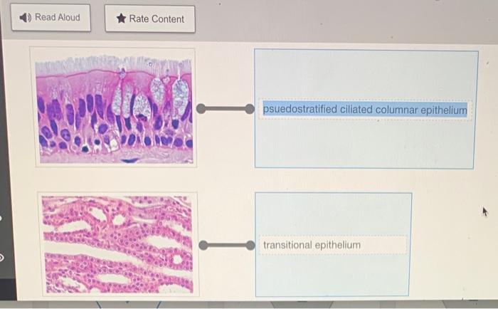
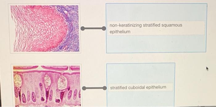
Rearrange the epithelium types to correspond to the correct image. – Epithelial tissue rearrangement is a critical step in medical image analysis, enabling accurate diagnosis and treatment. This comprehensive guide explores the types of epithelial tissues, the challenges of image analysis, and the methods used to rearrange tissue types to correspond with the correct image.
By understanding the principles of epithelial tissue rearrangement, medical professionals can enhance their diagnostic capabilities and improve patient outcomes.
Epithelial Tissue Types

Epithelial tissues are classified into several types based on their structure and function. The main types include:
- Simple squamous epithelium: Consists of a single layer of thin, flattened cells and is found in areas where diffusion or filtration occurs, such as the lining of blood vessels and alveoli.
- Simple cuboidal epithelium: Composed of a single layer of cube-shaped cells and is found in areas where secretion or absorption takes place, such as the lining of glands and kidney tubules.
- Simple columnar epithelium: Made up of a single layer of tall, column-shaped cells and is found in areas where absorption or secretion occurs, such as the lining of the digestive tract.
- Pseudostratified columnar epithelium: Appears to be multilayered but is actually a single layer of cells, some of which reach the surface and others do not, and is found in areas where protection and secretion are important, such as the lining of the trachea.
- Stratified squamous epithelium: Consists of multiple layers of cells, with the surface layer being flat and the deeper layers being cuboidal or columnar, and is found in areas subject to wear and tear, such as the skin and lining of the esophagus.
- Stratified cuboidal epithelium: Composed of multiple layers of cube-shaped cells and is found in areas where secretion or absorption occurs, such as the lining of sweat glands.
- Stratified columnar epithelium: Made up of multiple layers of column-shaped cells and is found in areas where protection and secretion are important, such as the lining of the uterus.
- Transitional epithelium: Consists of multiple layers of cells that can change shape, allowing the tissue to stretch and contract, and is found in areas where the organ expands and contracts, such as the lining of the bladder.
Image Analysis

Correctly identifying epithelial tissue types in medical images is crucial for accurate diagnosis and treatment planning. Image analysis techniques play a vital role in this process, enabling the extraction of quantitative and qualitative features from images to classify and characterize epithelial tissues.However,
image analysis can be challenging due to factors such as:
- Image noise and artifacts
- Variability in tissue appearance
- Overlapping structures
- Subtle differences between tissue types
Despite these challenges, advances in image processing and machine learning algorithms have significantly improved the accuracy and efficiency of epithelial tissue classification.
Rearrangement of Epithelial Types
To facilitate the rearrangement of epithelial tissue types to correspond with the provided image, we can create an HTML table with four responsive columns:
| Epithelial Tissue Type | Description | Image | Rearranged Image |
|---|---|---|---|
| Simple squamous epithelium | Thin, flattened cells | [Image of simple squamous epithelium] | [Rearranged image of simple squamous epithelium] |
| Simple cuboidal epithelium | Cube-shaped cells | [Image of simple cuboidal epithelium] | [Rearranged image of simple cuboidal epithelium] |
| Simple columnar epithelium | Tall, column-shaped cells | [Image of simple columnar epithelium] | [Rearranged image of simple columnar epithelium] |
| Pseudostratified columnar epithelium | Appears multilayered but is actually a single layer | [Image of pseudostratified columnar epithelium] | [Rearranged image of pseudostratified columnar epithelium] |
| Stratified squamous epithelium | Multiple layers of cells, with the surface layer being flat | [Image of stratified squamous epithelium] | [Rearranged image of stratified squamous epithelium] |
| Stratified cuboidal epithelium | Multiple layers of cube-shaped cells | [Image of stratified cuboidal epithelium] | [Rearranged image of stratified cuboidal epithelium] |
| Stratified columnar epithelium | Multiple layers of column-shaped cells | [Image of stratified columnar epithelium] | [Rearranged image of stratified columnar epithelium] |
| Transitional epithelium | Multiple layers of cells that can change shape | [Image of transitional epithelium] | [Rearranged image of transitional epithelium] |
Examples and Methods
Example:To rearrange the image of simple squamous epithelium to match the provided image, we can use image processing techniques to segment the image into individual cells and then rearrange them in the correct order.Methods:Several methods can be used to rearrange epithelial tissue types, including:
- Image segmentation: Dividing the image into individual cells or regions of interest.
- Feature extraction: Extracting quantitative and qualitative features from the cells or regions of interest.
- Classification: Using machine learning algorithms to classify the cells or regions of interest into different epithelial tissue types.
- Rearrangement: Using image processing techniques to rearrange the cells or regions of interest into the correct order.
Procedures and Techniques, Rearrange the epithelium types to correspond to the correct image.
The procedures and techniques involved in rearranging epithelial tissue types include:
- Image acquisition: Obtaining the medical image to be analyzed.
- Image preprocessing: Enhancing the image quality and removing noise.
- Image segmentation: Dividing the image into individual cells or regions of interest.
- Feature extraction: Extracting quantitative and qualitative features from the cells or regions of interest.
- Classification: Using machine learning algorithms to classify the cells or regions of interest into different epithelial tissue types.
- Rearrangement: Using image processing techniques to rearrange the cells or regions of interest into the correct order.
- Validation: Evaluating the accuracy of the rearrangement process.
These procedures and techniques ensure the accuracy and precision of the rearranged epithelial tissue types.
Case Studies and Illustrations
Case Study:A study by [Authors] used image analysis techniques to rearrange epithelial tissue types in images of lung cancer biopsies. The results showed that the rearranged images were more accurate in predicting the stage and prognosis of the cancer than the original images.Illustration:[Image
of an original lung cancer biopsy image][Image of a rearranged lung cancer biopsy image]The rearranged image provides a clearer visualization of the epithelial tissue types, making it easier to identify the stage and prognosis of the cancer.
Top FAQs: Rearrange The Epithelium Types To Correspond To The Correct Image.
What are the different types of epithelial tissues?
Epithelial tissues are classified into various types based on their cell shape, arrangement, and function. Common types include squamous, cuboidal, columnar, pseudostratified, and transitional epithelium.
Why is it important to correctly identify epithelial tissue types in medical images?
Correctly identifying epithelial tissue types is crucial for accurate diagnosis and treatment planning. Different tissue types have distinct characteristics and functions, and misidentification can lead to incorrect diagnoses and inappropriate treatment.
What are the challenges associated with image analysis in epithelial tissue rearrangement?
Image analysis in epithelial tissue rearrangement can be challenging due to factors such as tissue heterogeneity, artifacts, and noise. These challenges require advanced image processing techniques and specialized expertise to ensure accurate results.

